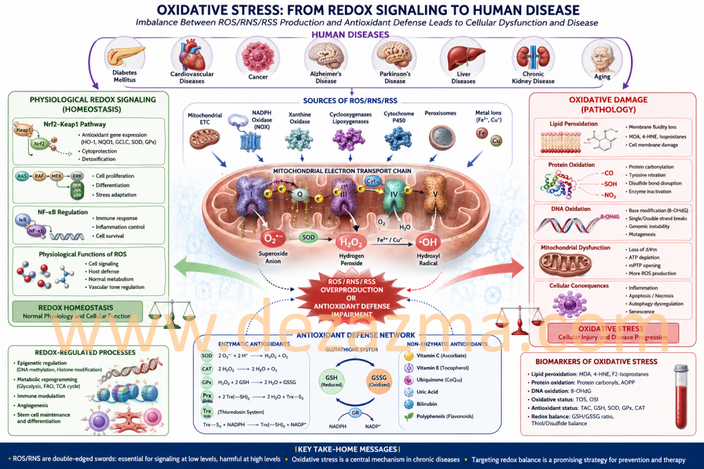
دزآزما

آموزش و پژوهش در بیوشیمی

استرس اکسیداتیو

31 می 26

استرس اکسیداتیو یکی از بنیادی‌ترین مفاهیم در بیوشیمی مدرن و زیست‌شناسی محسوب می‌شود و به وضعیتی اطلاق می‌گردد که در آن تعادل دینامیک میان تولید گونه‌های فعال اکسیژن، نیتروژن و گوگرد (Reactive Oxygen, Nitrogen and Sulfur Species; ROS/RNS/RSS) و ظرفیت مکانیسمهای آنتی‌اکسیدانی سلول مختل شده و محیط سلولی به سمت شرایط اکسیدکننده سوق می‌یابد.

برخلاف دیدگاه سنتی که ROS را صرفاً عوامل مخرب تلقی می‌کرد، امروزه مشخص شده است که این مولکول‌ها در غلظت‌های فیزیولوژیک به‌عنوان پیام‌رسان‌های ثانویه در تنظیم مسیرهای سیگنالینگ سلولی، تکثیر، تمایز، پاسخ ایمنی، تنظیم اپی‌ژنتیک، عملکرد میتوکندری و سازگاری متابولیک نقش اساسی ایفا می‌کنند. با این حال، افزایش کنترل‌نشده تولید ROS یا کاهش ظرفیت دفاع آنتی‌اکسیدانی منجر به آسیب تجمعی در سطح مولکولی، سلولی و بافتی می‌شود.

مهم‌ترین منابع درون‌سلولی تولید ROS شامل کمپلکس‌های I و III زنجیره انتقال الکترون میتوکندری، خانواده آنزیم‌های NADPH Oxidase (NOX)، زانتین اکسیداز، سیکلواکسیژنازها، لیپواکسیژنازها، سیستم سیتوکروم P450، پراکسی‌زوم‌ها و واکنش‌های وابسته به یون‌های فلزی واسطه مانند آهن و مس هستند.

در این میان، میتوکندری نه‌تنها منبع اصلی تولید سوپراکسید محسوب می‌شود، بلکه به‌عنوان هدف اولیه آسیب اکسیداتیو نیز شناخته می‌شود؛ به‌طوری‌که اختلال در عملکرد میتوکندری می‌تواند یک چرخه معیوب از افزایش تولید ROS، کاهش تولید ATP و تخریب ساختارهای سلولی را ایجاد کند.

در سطح مولکولی، استرس اکسیداتیو موجب پراکسیداسیون اسیدهای چرب غیراشباع غشایی و تولید متابولیت‌های واکنش‌پذیری نظیر مالون‌دی‌آلدئید (MDA)، 4-هیدروکسی‌نوننال (4-HNE) و ایزوپروستان‌ها می‌شود که خود دارای فعالیت‌های بیولوژیک و پاتوژنیک مستقل هستند. همچنین اکسیداسیون پروتئین‌ها منجر به تشکیل گروه‌های کربونیل، نیتروزیلاسیون تیروزین، اکسیداسیون باقی‌مانده‌های سیستئین و متیونین و تغییرات ساختاری برگشت‌ناپذیر می‌شود که عملکرد آنزیم‌ها، گیرنده‌ها و پروتئین‌های انتقال‌دهنده را مختل می‌کند. آسیب اکسیداتیو به DNA نیز شامل اکسیداسیون بازهای نوکلئوتیدی، تشکیل 8-هیدروکسی-2′-دئوکسی‌گوانوزین (8-OHdG)، شکست رشته‌های DNA و افزایش ناپایداری ژنومی است که در بروز سرطان و فرآیند پیری نقش محوری دارند.

در پاسخ به این چالش، سلول‌ها شبکه‌ای پیچیده از مکانیسمهای دفاعی را توسعه داده‌اند که شامل آنتی‌اکسیدان‌های آنزیمی نظیر سوپراکسید دیسموتاز (SOD)، کاتالاز (CAT)، گلوتاتیون پراکسیداز (GPx)، پراکسی‌ردوکسین‌ها (Prx) و تیوردوکسین‌ها (Trx) و نیز آنتی‌اکسیدان‌های غیرآنزیمی همچون گلوتاتیون احیاشده (GSH)، کوآنزیم Q10، ویتامین C، ویتامین E، اسید اوریک، بیلی‌روبین و پلی‌فنول‌های گیاهی است. تنظیم بیان بسیاری از این سیستم‌های دفاعی توسط فاکتور رونویسی Nrf2 انجام می‌شود که امروزه یکی از مهم‌ترین اهداف درمانی در پزشکی مولکولی محسوب می‌گردد.

از منظر بالینی، شواهد گسترده نشان می‌دهد که استرس اکسیداتیو در پاتوژنز طیف وسیعی از بیماری‌ها از جمله دیابت ملیتوس، سندرم متابولیک، آترواسکلروز، بیماری‌های قلبی–عروقی، سرطان، بیماری آلزایمر، پارکینسون، بیماری‌های خودایمنی، نارسایی کلیوی، بیماری‌های کبدی و اختلالات مرتبط با سالمندی نقش اساسی دارد. علاوه بر این، تعامل پیچیده میان استرس اکسیداتیو، التهاب مزمن، اختلال عملکرد میتوکندری و تغییرات اپی‌ژنتیک به‌عنوان یک محور مرکزی در زیست‌شناسی بیماری‌های مزمن شناخته می‌شود.

روشهای سنجش قدرت آنتی اکسیدان

16 مارس 10

ANTIOXIDANT ASSAYS

From Oxford Biomedical Research: http://www.oxfordbiomed.com/commerce/info/showpage.jsp?page_id=1042&gclid=CL603tWq15kCFRpN5QodpDEpVw/

Background
It is now well established that oxidative stress is a major risk factor for the development of several diseases including atherosclerosis, cardiovascular disease, and cancer. Oxidative stress is the condition in which there is an imbalance between the concentrations of reactive oxygen species (ROS) and physiological antioxidants, resulting in oxidative damage to many biomolecules within the cell. Products of ROS-mediated oxidation are widely used to monitor oxidative stress. However, it is also important to assess the antioxidant capacity of cells and biological fluids, as well as putative “functional foods” to assess their antioxidant capacity. Organisms possess multiple antioxidant systems to help regulate ROS and prevent oxidative stress. In vertebrates, these include enzymes that metabolize ROS, antioxidant proteins, and smaller molecules that are important antioxidants. These antioxidants include hydrophilic as well as lipid-soluble molecules that are localized throughout various tissues and cell types.

Given the multiplicity of antioxidant pathways, their centrality in the prevention of oxidative stress, and the influences of lifestyle and nutritional supplements on an individual’s antioxidant capacity, it is important to be able to quantitatively measure the total antioxidant capacity or antioxidant power in a biological specimen or in nutrients.

ORAC Assay

Assay Principle
The ORAC assay is based on the oxidation of fluorescein by peroxyl radicals via a classic hydrogen atom transfer (HAT) mechanism. Free radicals are generated by the water soluble compound 2,2’-azobis-2-methyl-propanimidamide (AAPH). The peroxyl radicals thus generated quench the fluorescence of fluorescein over time. The antioxidants block the peroxyl radical mediated oxidation of fluorescein until all of the antioxidant activity in the sample is exhausted, after which the AAPH-generated peroxyl radicals react with and quench the fluorescence of fluorescein. The area under the fluorescence decay curve (AUC) is used to quantify the total peroxyl radical antioxidant activity in a sample and is compared to a standard curve obtained using various concentrations of the water soluble vitamin E analog Trolox. Unlike other antioxidant activity assays, the fluorescent ORAC assay provides a direct measurement of antioxidant capacity against hydrophilic chain-breaking peroxyl radicals.

HORAC Assay

Assay Principle
The HORAC assay is based on the oxidation of fluorescein by hydroxyl radicals via a classic hydrogen atom transfer (HAT) mechanism. Free radicals are generated by hydrogen peroxide (H2O2). The hydroxyl radicals thus generated quench the fluorescence of fluorescein over time. The antioxidants block the hydroxyl radical mediated oxidation of fluorescein until all of the antioxidant activity in the sample is exhausted, after which the H2O2 radicals react with and quench the fluorescence of fluorescein. The area under the fluorescence decay curve (AUC) is used to quantify the total hydroxyl radical antioxidant activity in a sample and is compared to a standard curve obtained using various concentrations of gallic acid. Unlike other antioxidant activity assays, the fluorescent HORAC assay provides a direct measurement of antioxidant capacity against hydrophilic chain-breaking hydroxyl radicals.

Total Antioxidant Power

Background
Oxidative stress has been implicated in a number of diseases such as atherosclerosis, chronic inflammatory disease, chronic renal failure, and cancer.  It is a condition where an imbalance exists between the production of reactive oxidizing species and the body’s ability to neutralize these intermediates, resulting in cellular damage. The body has designed several physiological responses to oxidative stress including counterbalances such as enzymes and variously functionalized molecules (see examples below) that effectively neutralize these damaging species. These antioxidants can be either water or lipid soluble, and are localized transiently throughout various tissues, cells and cell types.

Given the multiplicity of antioxidant pathways, their centrality in the prevention of oxidant stress, and the influences of lifestyle and nutritional supplements on an individual’s antioxidant capacity, it is important to be able to quantitatively measure the total antioxidant capacity or antioxidant power with biological specimens.

Assay Principles
The reduction potential of the sample or standard effectively converts Cu+2 to Cu+1, thus changing the ion’s absorption characteristics. This reduced form of copper will selectively form a stable 2:1 complex with the chromogenic reagent with an absorption maximum at ca. 450 nm. A known concentration of uric acid is used to create a calibration curve, with the data being expressed as mM uric acid equivalents or in μM copper reducing equivalents