دزآزما

آموزش و پژوهش در بیوشیمی

Cellular Sources and Spatial Organization of Reactive Species: An Integrated Redox Architecture

9 ژوئن 26

1. Overview of Enzymatic ROS/RNS/RSS Generation

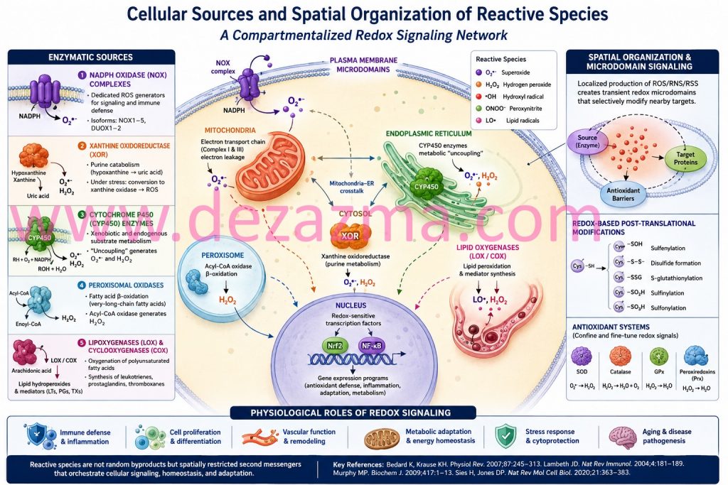
Reactive oxygen, nitrogen, and sulfur species (ROS, RNS, RSS) emerge from a highly integrated enzymatic network distributed across multiple subcellular compartments. Rather than representing stochastic byproducts of metabolism and oxidative stress, these species are generated through specialized redox circuits that function as signaling modules within cellular physiology. Each enzymatic system contributes distinct kinetic and spatial characteristics to intracellular redox dynamics, collectively forming a hierarchical oxidative signaling architecture.¹–³

2. NADPH Oxidase (NOX) Complexes: Dedicated Redox Signal Transducers

NADPH oxidase (NOX) complexes constitute prototypical “professional ROS-generating systems” whose primary biological role is signal transduction rather than energy metabolism. These multi-subunit flavocytochrome assemblies catalyze electron transfer from NADPH to molecular oxygen, producing superoxide (O₂•⁻) as a regulated signaling intermediate.⁴,⁵ Isoform-specific localization (NOX1–5, DUOX1–2) enables compartmentalized ROS generation in the plasma membrane, endosomal membranes, and specialized secretory vesicles.

NOX-derived ROS function as tightly regulated redox bursts that orchestrate immune defense, phagocytic microbial killing, endothelial activation, and growth factor signaling. Importantly, these signals exhibit high spatial fidelity, ensuring that oxidative modifications remain confined to proximal target proteins.⁵,⁶

3. Xanthine Oxidoreductase: Metabolic–Redox Interface Enzyme

Xanthine oxidoreductase (XOR) operates as a bidirectional metabolic–redox switch within purine degradation pathways. Under physiological conditions, XOR predominantly exists as xanthine dehydrogenase; however, proteolytic conversion or oxidative modification shifts enzymatic behavior toward xanthine oxidase, a potent ROS-producing form.⁷

This conversion results in enhanced electron leakage to molecular oxygen, generating superoxide and hydrogen peroxide. XOR-derived ROS become particularly relevant in ischemia–reperfusion injury, where substrate accumulation and oxygen reintroduction converge to produce oxidative bursts that amplify endothelial dysfunction and inflammatory signaling cascades.⁷,⁸

4. Cytochrome P450 Systems: Redox Leakage in Xenobiotic Metabolism

Cytochrome P450 (CYP) monooxygenases represent a vast superfamily of heme-thiolate enzymes localized predominantly in the endoplasmic reticulum. Their canonical role in xenobiotic detoxification and steroid biosynthesis is accompanied by intrinsic electron uncoupling; wherein incomplete reduction of oxygen generates superoxide and hydrogen peroxide as byproducts.⁹

This redox leakage is particularly pronounced under conditions of substrate saturation or metabolic stress, linking hepatic detoxification processes with intracellular oxidative burden. CYP-derived ROS also contribute to lipid peroxidation cascades and electrophilic stress formation.⁹,¹⁰

5. Peroxisomal Oxidases: Compartmentalized Hydrogen Peroxide Production

Peroxisomes function as specialized oxidative organelles enriched in flavin-dependent oxidases that generate hydrogen peroxide as a primary metabolic product. Acyl-CoA oxidase-mediated β-oxidation of very-long-chain fatty acids represents a central source of peroxisomal H₂O₂ production.¹¹

Unlike mitochondrial ATP-coupled oxidation, peroxisomal metabolism decouples electron transfer from energy conservation, resulting in obligatory ROS formation. Catalase and peroxiredoxin systems tightly regulate peroxisomal redox output, preventing cytotoxic accumulation while enabling localized signaling functions.¹¹,¹²

6. Lipoxygenases and Cyclooxygenases: Lipid Radical Chemistry in Inflammatory Signaling

Lipoxygenases (LOX) and cyclooxygenases (COX) catalyze oxygenation of polyunsaturated fatty acids via radical-mediated mechanisms that intrinsically involve reactive oxygen intermediates. These enzymes generate lipid hydroperoxides and prostanoid precursors that serve as bioactive mediators of inflammation, immunity, and vascular tone regulation.¹³

LOX-derived leukotrienes and COX-derived prostaglandins function as both downstream effectors and amplifiers of redox signaling. Their biosynthesis integrates oxidative chemistry with immunological programming, positioning lipid peroxidation pathways as central nodes in inflammation-associated redox networks.¹³,¹⁴

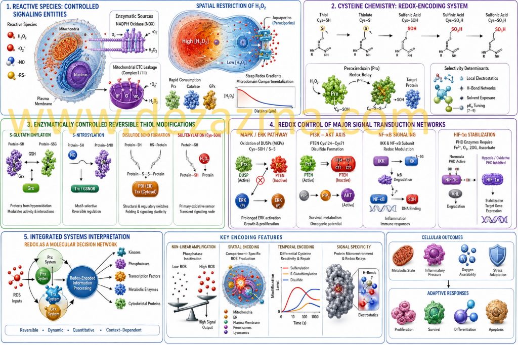
7. Spatial Compartmentalization of Redox Signaling

A defining principle of modern redox biology is the spatial restriction of reactive species generation within subcellular microdomains. Mitochondria, endoplasmic reticulum, peroxisomes, nucleus, and plasma membrane nanoregions each maintain distinct redox microenvironments governed by localized enzymatic production and antioxidant buffering systems.¹,²

This compartmentalization ensures that ROS act as short-range second messengers rather than diffusible cytotoxins. Within these microdomains, reversible oxidation of cysteine and selenocysteine residues modulates protein conformation, enzymatic activity, and transcriptional output.³

References

Sies H. Oxidative stress: a concept in redox biology and medicine. Redox Biol. 2015;4:180–183.

Holmström KM, Finkel T. Cellular mechanisms and physiological consequences of redox-dependent signaling. Nat Rev Mol Cell Biol. 2014;15(6):411–421.

Jones DP, Sies H. The redox code. Antioxid Redox Signal. 2015;23(9):734–746.

Bedard K, Krause KH. The NOX family of ROS-generating NADPH oxidases: physiology and pathophysiology. Physiol Rev. 2007;87(1):245–313.

Lambeth JD. NOX enzymes and the biology of reactive oxygen. Nat Rev Immunol. 2004;4(3):181–189.

Ushio-Fukai M. Compartmentalization of redox signaling through NOX family NADPH oxidases. Free Radic Biol Med. 2009;46(2):129–141.

Pacher P, Nivorozhkin A, Szabó C. Therapeutic effects of xanthine oxidase inhibitors. Pharmacol Rev. 2006;58(1):87–114.

Granger DN, Kvietys PR. Reperfusion injury and reactive oxygen species. J Physiol. 2015;593(2):341–348.

Zangar RC, Davydov DR, Verma S. Mechanisms that regulate cytochrome P450. Toxicol Appl Pharmacol. 2004;199(3):316–331.

Guengerich FP. Cytochrome P450 and chemical toxicology. Chem Res Toxicol. 2008;21(1):70–83.

Wanders RJA, Waterham HR. Biochemistry of mammalian peroxisomes. Annu Rev Biochem. 2006;75:295–332.

Fransen M, Nordgren M, Wang B, Apanasets O. Role of peroxisomes in ROS metabolism. Antioxid Redox Signal. 2012;16(9):1047–1058.

Dennis EA, Norris PC. Eicosanoid storm in infection and inflammation. Nat Rev Immunol. 2015;15(8):511–523.

Ricciotti E, FitzGerald GA. Prostaglandins and inflammation. Arterioscler Thromb Vasc Biol. 2011;31(5):986–1000.

Redox Signaling and Molecular Switching Mechanisms: A Systems-Level and Chemical Biology Perspective

9 ژوئن 26

1. Reactive Species as Spatially and Temporally Controlled Signaling Entities

Reactive oxygen, nitrogen, and sulfur species (ROS/RNS/RSS) are increasingly understood as tightly regulated signaling mediators embedded within compartmentalized enzymatic networks rather than stochastic byproducts of oxidative metabolism. Hydrogen peroxide (H₂O₂), in particular, exhibits a unique physicochemical profile: moderate reactivity, relative stability compared to radical species, and selective oxidation of low pKa cysteine thiolates (Cys–S⁻), enabling signaling specificity under controlled microenvironmental conditions [1–3].

A defining principle of redox signaling is spatial restriction. H₂O₂ diffusion is not purely passive but is shaped by membrane permeability, aquaporin-mediated transport (peroxiporins), and rapid enzymatic consumption by peroxiredoxins (Prx), catalase, and glutathione peroxidases (GPx). This establishes steep intracellular redox gradients, particularly between mitochondria, endoplasmic reticulum, and cytosolic microdomains [4–6].

At the enzymatic source level, NADPH oxidase (NOX) complexes generate ROS in a regulated, receptor-coupled manner, while mitochondrial electron transport chain leakage (Complex I/III) contributes to basal redox tone. Importantly, these sources are not redundant but functionally specialized, producing discrete signaling pulses with distinct subcellular localization and temporal kinetics [7,8].

2. Cysteine Chemistry as a Redox-Encoding System

Protein cysteine residues operate as chemically programmable sensors due to their tunable pKa (typically 7–9), nucleophilicity, and susceptibility to stepwise oxidation states. The redox transition landscape includes:

Among these, sulfenylation (–SOH) is a critical kinetic intermediate, functioning as a transient signaling node rather than damage marker [9,10].

Thiol (Cys–SH) → thiolate (Cys–S⁻)

Sulfenic acid (Cys–SOH)

Sulfinic acid (Cys–SO₂H, partially reversible via sulfiredoxin in select proteins)

Sulfonic acid (Cys–SO₃H, generally irreversible physiological endpoint)

Selectivity in cysteine oxidation is not determined solely by solvent exposure but by local electrostatics, hydrogen bonding networks, and proximity to catalytic redox relays such as peroxiredoxins. In fact, peroxiredoxins function not only as antioxidants but also as H₂O₂ sensors and signal transducers via redox relay mechanisms, transferring oxidative equivalents to target proteins with high specificity [11,12].

3. Enzymatically Controlled Reversible Thiol Modifications

Redox-based post-translational modifications form an interconnected regulatory system:

S-glutathionylation arises through thiol–disulfide exchange or via sulfenic acid intermediates reacting with reduced glutathione (GSH). This modification protects cysteine residues from hyperoxidation while simultaneously altering protein conformation, enzymatic activity, and protein–protein interactions. It is dynamically regulated by glutaredoxin (Grx) systems [13].

S-nitrosylation is mediated through NO-derived nitrosating equivalents (e.g., N₂O₃, metal–NO complexes) and exhibits motif selectivity influenced by acid–base microenvironments and hydrophobic pockets. Denitrosylation is enzymatically regulated by thioredoxin and S-nitrosoglutathione reductase (GSNOR), enabling reversibility [14].

Disulfide bond formation acts as both structural and regulatory switches. In signaling contexts, dynamic disulfide exchange reactions are catalyzed by protein disulfide isomerases (PDI) in the ER and thioredoxin systems in the cytosol, ensuring controlled folding and signaling plasticity [15].

Sulfenylation (Cys–SOH) represents the primary oxidative sensing event. Its detection in living systems has revealed that many proteins undergo rapid, reversible sulfenylation bursts within seconds of ROS exposure, consistent with signal transduction rather than oxidative damage [16].

4. Redox Control of Major Signal Transduction Networks

MAPK/ERK Pathway

Redox regulation of MAPK signaling is primarily mediated through oxidation-sensitive phosphatases, particularly dual-specificity phosphatases (DUSPs/MKPs). Oxidation of catalytic cysteine residues within the active site (forming sulfenyl or disulfide states) inactivates phosphatase activity, thereby prolonging ERK phosphorylation and amplifying proliferative signaling outputs. This mechanism introduces a redox-dependent “gain control” layer over growth factor signaling [17].

PI3K–AKT Axis

The tumor suppressor PTEN contains a catalytically essential cysteine (Cys124) highly susceptible to reversible oxidation. Formation of a disulfide bond between Cys124 and Cys71 leads to conformational inactivation of PTEN phosphatase activity, resulting in increased PIP₃ accumulation and downstream AKT activation. This redox switch directly links oxidative tone to metabolic reprogramming, survival signaling, and oncogenic transformation potential [18].

NF-κB Signaling

NF-κB regulation exhibits multilayered redox sensitivity. At the upstream level, IKK complex activation is modulated by thiol oxidation and S-glutathionylation. At the transcriptional level, cysteine residues within the DNA-binding domain of NF-κB subunits (e.g., p50 Cys62) undergo redox modifications that alter DNA affinity. Thus, ROS functions both as an activator and modulator of transcriptional specificity depending on intensity and duration [19].

HIF-1α Stabilization Pathway

Hypoxia-inducible factor regulation is tightly coupled to redox state via prolyl hydroxylase domain (PHD) enzymes, which require Fe²⁺ and O₂ for activity. Oxidative modulation of iron center chemistry, as well as ascorbate and 2-oxoglutarate availability, suppresses hydroxylation of HIF-1α, preventing VHL-mediated degradation. This integrates oxygen sensing with redox signaling, allowing metabolic adaptation under hypoxic and oxidative stress conditions [20].

5. Integrated Systems Interpretation: Redox as a Molecular Decision Network

At a systems level, redox signaling constitutes a chemically encoded information-processing layer superimposed on classical phosphorylation networks.

The essential features include:

Non-linear amplification via reversible enzyme inactivation (phosphatases > kinases)

Spatial encoding via compartment-specific ROS production

Temporal encoding via differential cysteine reactivity and repair kinetics

Signal specificity via protein microenvironment and redox relays

Peroxiredoxin-based signal relays and thioredoxin/glutathione systems form a hierarchical buffering architecture that transforms oxidative inputs into graded, reversible signaling outputs. This enables cells to interpret redox fluctuations as quantitative parameters reflecting metabolic state, inflammatory pressure, and oxygen availability rather than binary stress signals [21,22].

References

Sies H. Hydrogen peroxide as a central redox signaling molecule. Redox Biol. 2017;11:613–619.

D’Autréaux B, Toledano MB. ROS signaling specificity. Nat Rev Mol Cell Biol. 2007;8:813–824.

Winterbourn CC. The biological chemistry of hydrogen peroxide. Methods Enzymol. 2013;528:3–25.

Rhee SG. Redox signaling: H₂O₂ as intracellular messenger. Exp Mol Med. 1999;31:53–59.

Miller EW et al. Aquaporin-mediated H₂O₂ transport. PNAS. 2010;107:15681–15686.

Veal EA et al. Peroxiredoxins as H₂O₂ sensors. J Biol Chem. 2007;282:26753–26759.

Bedard K, Krause KH. NOX family NADPH oxidases. Physiol Rev. 2007;87:245–313.

Murphy MP. Mitochondrial ROS signaling. Biochem J. 2009;417:1–13.

Poole LB, Nelson KJ. Cysteine oxidation mechanisms. Curr Opin Chem Biol. 2008;12:18–23.

Reddie KG, Carroll KS. Protein cysteine sulfenylation. Curr Opin Chem Biol. 2008;12:746–754.

Wood ZA et al. Peroxiredoxin catalytic cycle. Trends Biochem Sci. 2003;28:32–40.

Sobotta MC et al. Peroxiredoxin redox relays. PNAS. 2015;112:106–111.

Giustarini D et al. S-glutathionylation in signaling. Antioxid Redox Signal. 2004;6:345–360.

Foster MW et al. S-nitrosylation signaling. Trends Mol Med. 2009;15:391–404.

Hatahet F, Ruddock LW. Protein disulfide isomerase systems. FEBS J. 2009;276:194–201.

Paulsen CE, Carroll KS. Sulfenic acid biology. Chem Rev. 2013;113:4633–4679.

Kamata H et al. Redox regulation of MAPK signaling. Cell Signal. 1999;11:1–14.

Leslie NR. PTEN oxidation and PI3K signaling. Antioxid Redox Signal. 2007;9:1765–1774.

Morgan MJ, Liu ZG. ROS and NF-κB crosstalk. Cell Res. 2011;21:103–115.

Kaelin WG, Ratcliffe PJ. Oxygen sensing via HIF. Mol Cell. 2008;30:393–402.

Winterbourn CC. Redox biology complexity. Biochem J. 2013;461:1–10.

Jones DP. Redox sensing and signaling. Nat Rev Mol Cell Biol. 2006;7:392–401.

Oxidative Stress

7 ژوئن 26

1. Overview: From Damage Concept to Redox Regulatory Network

Oxidative stress is a central concept in modern molecular and cellular biology, describing a disruption in redox homeostasis caused by an imbalance between the generation of reactive species and the capacity of antioxidant systems. Contemporary definitions extend beyond the classical “oxidative damage” model and recognize reactive oxygen species (ROS), reactive nitrogen species (RNS), and reactive sulfur species (RSS) as integral components of cellular physiology.

In biological systems, redox balance is not static but dynamically regulated. Cells operate under a finely tuned equilibrium in which reactive species function as signaling intermediates at low to moderate concentrations, while excessive accumulation leads to macromolecular damage and functional disruption. This dual role has led to the conceptual refinement of oxidative stress into oxidative eustress (physiological signaling) and oxidative distress (pathological overload).

In biological systems, redox balance is not static but dynamically regulated. Cells operate under a finely tuned equilibrium in which reactive species function as signaling intermediates at low to moderate concentrations, while excessive accumulation leads to macromolecular damage and functional disruption. This dual role has led to the conceptual refinement of oxidative stress into oxidative eustress (physiological signaling) and oxidative distress (pathological overload).

In biological systems, redox balance is not static but dynamically regulated. Cells operate under a finely tuned equilibrium in which reactive species function as signaling intermediates at low to moderate concentrations, while excessive accumulation leads to macromolecular damage and functional disruption. This dual role has led to the conceptual refinement of oxidative stress into oxidative eustress (physiological signaling) and oxidative distress (pathological overload).

2. Cellular Sources and Spatial Organization of Reactive Species

Reactive species are generated through multiple enzymatic and non-enzymatic processes distributed across cellular compartments. The most prominent intracellular source is the mitochondrial electron transport chain (ETC), particularly Complex I and Complex III, where electron leakage to molecular oxygen produces superoxide (O₂•⁻).

However, mitochondria represent only one node in a broader redox network. Additional enzymatic sources include:

• NADPH oxidase (NOX) complexes, which deliberately generate ROS for signaling and immune defense

• Xanthine oxidoreductase, involved in purine metabolism

• Cytochrome P450 enzymes, participating in xenobiotic metabolism

• Peroxisomal oxidases, contributing to fatty acid oxidation

• Lipoxygenases and cyclooxygenases, involved in lipid mediator synthesis

Importantly, ROS production is spatially compartmentalized. Distinct subcellular microenvironments (mitochondria, endoplasmic reticulum, nucleus, and plasma membrane) generate localized redox signals. This spatial restriction ensures specificity in redox signaling and prevents uncontrolled diffusion of reactive species.

3. Redox Signaling and Molecular Switching Mechanisms

Contrary to earlier assumptions, reactive species are not merely toxic byproducts but act as second messengers in intracellular signaling pathways. Among them, hydrogen peroxide (H₂O₂) plays a dominant role due to its relative stability and ability to diffuse across membranes via aquaporins.

Redox signaling operates primarily through reversible oxidation of cysteine residues in target proteins. These thiol-based modifications function as molecular switches regulating protein activity. Key reversible modifications include:

• S-glutathionylation

• S-nitrosylation

• Disulfide bond formation

• Sulfenylation (–SOH formation)

These modifications regulate signaling cascades such as:

• MAPK/ERK pathways (cell proliferation)

• PI3K–AKT signaling (metabolic regulation)

• NF-κB activation (immune and inflammatory responses)

• HIF-1α stabilization (hypoxic adaptation)

Thus, redox signaling integrates environmental cues into cellular decision-making processes.

4. Mitohormesis and Adaptive Stress Responses

A key concept in modern redox biology is hormesis, particularly mitohormesis, where low levels of mitochondrial ROS induce adaptive stress resistance. Instead of causing damage, mild oxidative stress activates protective pathways that enhance cellular fitness.

Mitohormetic responses include:

• Upregulation of antioxidant enzymes (SOD, CAT, GPx)

• Activation of mitochondrial biogenesis (via PGC-1α)

• Enhanced DNA repair capacity

• Improved proteostasis and autophagy

This adaptive framework explains why physiological stressors such as exercise, caloric restriction, and intermittent metabolic stress can enhance cellular resilience and extend functional lifespan.

5. Mitochondria as Signaling Hubs and Retrograde Communication Centers

Mitochondria occupy a dual role as both major sources and sensitive targets of oxidative stress. Beyond ATP production, mitochondria function as signaling organelles that communicate their functional state to the nucleus through mitochondrial retrograde signaling.

When mitochondrial function is altered, ROS act as signaling mediators that modify nuclear gene expression. This leads to metabolic reprogramming, including:

• Shift toward glycolytic metabolism under stress

• Induction of antioxidant gene networks

• Regulation of apoptosis and survival pathways

Mitochondrial dysfunction also amplifies ROS production, creating a self-reinforcing loop of oxidative amplification known as the “vicious cycle” of mitochondrial oxidative injury.

6. Oxidative Stress and Proteostasis Networks

Cellular protein homeostasis (proteostasis) is tightly regulated by redox mechanisms. Oxidative modifications influence protein folding, stability, and degradation.

Major proteostatic systems affected by ROS include:

• Molecular chaperones (heat shock proteins)

• Ubiquitin–proteasome system (UPS)

• Autophagy and selective mitophagy

Oxidative damage can lead to protein misfolding, aggregation, and loss of function. However, moderate ROS levels also activate proteostatic pathways, indicating a regulatory rather than purely destructive role.

Failure of proteostasis is a key feature of aging and neurodegenerative processes.

7. Redox Regulation of Transcriptional Networks

Gene expression is extensively controlled by redox-sensitive transcription factors. These include:

• Nrf2 (Nuclear factor erythroid 2–related factor 2): master regulator of antioxidant response

• NF-κB: immune and inflammatory signaling

• FOXO proteins: stress resistance and longevity regulation

• HIF-1α: hypoxic adaptation

• AP-1: proliferation and differentiation

• p53: DNA damage response and apoptosis

Nrf2 is particularly important because it coordinates the expression of detoxifying enzymes, glutathione metabolism genes, and cytoprotective proteins. Under oxidative stress, Nrf2 translocates to the nucleus and activates antioxidant response elements (AREs), establishing a broad defensive transcriptional program.

8. Oxidative DNA Damage and Genome Stability

Reactive species interact directly with nucleic acids, producing oxidative lesions such as base modifications, strand breaks, and crosslinking. A widely recognized marker of oxidative DNA damage is 8-hydroxy-2′-deoxyguanosine (8-OHdG).

Persistent oxidative DNA damage contributes to:

• Mutagenesis

• Genomic instability

• Altered gene expression

• Cellular senescence

DNA repair pathways, including base excision repair (BER), are essential for maintaining genomic integrity under oxidative conditions.

9. Epigenetic and Systems-Level Redox Regulation

Beyond direct molecular damage, oxidative stress influences epigenetic regulation, including:

• DNA methylation patterns

• Histone modifications

• Chromatin remodeling

• Non-coding RNA expression

These changes produce long-term alterations in gene expression profiles, linking environmental stress exposure to stable cellular phenotypes.

At the systems level, redox biology operates as an integrated network in which metabolism, signaling, gene regulation, and organelle function are interconnected. This perspective has led to the development of redox systems biology, which views oxidative stress as a network property rather than a single molecular event.

10. Conclusion: Oxidative Stress as a Biological Regulatory Principle

Oxidative stress is no longer understood as a purely pathological condition but as a fundamental regulatory principle in biology. Reactive species serve as essential mediators of cellular signaling, metabolic adaptation, and environmental responsiveness. Cellular outcomes depend on the intensity, duration, and spatial distribution of redox signals.

At physiological levels, redox signaling supports homeostasis, adaptation, and survival. At excessive levels, oxidative distress disrupts biomolecular integrity and cellular function. The balance between these states is maintained through complex antioxidant systems, transcriptional regulators such as Nrf2, and adaptive mechanisms including mitohormesis and proteostasis networks.

Understanding oxidative stress as an integrated redox signaling system provides a unified framework linking molecular biology, cell physiology, and systems-level adaptation in living organisms.

Core Concepts of the Article

1. Oxidative Stress

Central conceptual hub of redox biology and cellular regulation.

2. Reactive Oxygen Species (ROS) in Cells

Core molecular basis of redox biology; very high academic search volume.

3. Antioxidant Systems in Cells

Enzymatic and non-enzymatic defense mechanisms in cellular biology.

4. Mitochondrial ROS and Bioenergetics

Links respiration, electron transport chain, and redox signaling.

5. Redox Signaling and Cellular Communication

Focus on signaling networks rather than pathology.

6. Redox Homeostasis in Biological Systems

Systems biology perspective of balance and regulation.

7. Compartmentalized ROS Production

Advanced concept: spatial redox biology (mitochondria, ER, peroxisomes, membrane).

8. Thiol-Based Redox Regulation (Cysteine Biology)

Protein-level redox switching and molecular regulation.

9. Oxidative Stress and Cellular Adaptation

Includes hormesis, stress responses, and environmental adaptation.

10. Oxidative DNA Damage and Repair Mechanisms

Focus on molecular biology (mutation, repair systems, genome stability) without clinical framing.

Oxidative Stress Flashcards For Clinicians, Biologists and Laboratory Technicians

6 ژوئن 26

Some ideas for your future studies

Oxidative stress

Mitochondrial ROS and disease mechanisms

Clinical biomarkers of oxidative stress MDA, 8-OHdG, and isoprostanes

Oxidative stress in cardiovascular disease mechanisms and pathways

Role of oxidative stress in Alzheimer and Parkinson

Laboratory methods for oxidative stress biomarkers LC-MS MS HPLC

Redox biology and oxidative stress in human disease

NRF2 pathway role in oxidative stress and inflammation control

Lipid peroxidation and cellular membrane damage mechanisms

Ferroptosis and iron-driven oxidative cell death in disease

Oxidative stress and inflammation in chronic disease

Mitochondrial dysfunction and reactive oxygen species in pathology

Oxidative DNA damage biomarkers and clinical significance 8-OHdG

Antioxidant defense systems SOD, catalase, and glutathione peroxidase

Oxidative stress in diabetes complications and microvascular damage

Systems biology view of oxidative stress and redox signaling networks

Clinical interpretation of oxidative stress laboratory results explained

Comprehensive guide to oxidative stress disease mechanisms and testing

Oxidative Stress Map

6 ژوئن 26
OS

?What is Oxidative stress

6 ژوئن 26

Oxidative stress is a central concept in modern molecular and cellular biology, describing a disruption in redox homeostasis caused by an imbalance between the generation of reactive species and the capacity of antioxidant systems. Contemporary definitions extend beyond the classical “oxidative damage” model and recognize reactive oxygen species (ROS), reactive nitrogen species (RNS), and reactive sulfur species (RSS) as integral components of cellular physiology.

In biological systems, redox balance is not static but dynamically regulated. Cells operate under a finely tuned equilibrium in which reactive species function as signaling intermediates at low to moderate concentrations, while excessive accumulation leads to macromolecular damage and functional disruption. This dual role has led to the conceptual refinement of oxidative stress into oxidative eustress (physiological signaling) and oxidative distress (pathological overload).

Some useful concepts

What is oxidative stress

Oxidative stress explained for medical students

Reactive oxygen species (ROS) and antioxidant defense

Nrf2 signaling pathway and oxidative stress

Role of glutathione in oxidative stress

Oxidative stress biomarkers in clinical practice

Mitochondrial dysfunction and oxidative stress

Oxidative DNA damage and DNA repair

Some useful keywords for your future studies:

Oxidative Stress: Complete Guide

Sources of Reactive Oxygen Species

Causes of Oxidative Stress

Physiological vs Pathological ROS

Oxidative Stress and Aging

Oxidative Stress Biomarkers

Reactive Oxygen Species (ROS) and Redox Biology

Superoxide Radical

Hydrogen Peroxide

Hydroxyl Radical

Reactive Nitrogen Species

Reactive Sulfur Species

Redox Homeostasis

Antioxidant Defense Systems

Superoxide Dismutase

Catalase

Glutathione Peroxidase

Glutathione System

Vitamin C

Vitamin E

Dietary Antioxidants

Nrf2 Signaling Pathway

Nrf2-Keap1 Mechanism

Antioxidant Response Elements

Nrf2 in Cancer

Nrf2 in Neurodegeneration

Pharmacological Nrf2 Activators

Mitochondrial Dysfunction and Oxidative Stress

Electron Transport Chain ROS

Mitochondrial DNA Damage

Mitochondrial Aging

Mitophagy

Mitochondrial Diseases

Oxidative Stress Biomarkers

Malondialdehyde (MDA)

4-Hydroxynonenal (4-HNE)

8-OHdG

Total Antioxidant Capacity

Clinical Laboratory Testing

Oxidative Stress and Inflammation

NF-κB Signaling

Cytokines and ROS

Chronic Inflammation

Sepsis and Oxidative Damage

Immune Regulation

Oxidative DNA Damage and Genomic Instability

8-Oxo-Guanine

DNA Repair Mechanisms

Base Excision Repair

Mutagenesis

Cancer Initiation

Oxidative Stress in Human Disease

Cancer

Diabetes

Cardiovascular Disease

Alzheimer’s Disease

Parkinson’s Disease

Chronic Kidney Disease

Oxidative Stress in Microbiology and Host–Pathogen Interactions

NADPH Oxidase

Respiratory Burst

Chronic Granulomatous Disease

Bacterial Antioxidant Enzymes

ROS in Infection

Ferroptosis and Regulated Cell Death

GPX4

Iron Metabolism

Lipid Peroxidation

Ferroptosis in Cancer

  Therapeutic Targets

Some Useful phrases
Oxidative Stress
Central concept in redox biology and cellular regulation.
Reactive Oxygen Species (ROS) in Cells
Core molecular basis of redox biology
Antioxidant Systems in Cells
Enzymatic and non-enzymatic defense mechanisms in cellular biology.
Mitochondrial ROS and Bioenergetics
Links respiration, electron transport chain, and redox signaling.
Redox Signaling and Cellular Communication
Focus on signaling networks rather than pathology.
Redox Homeostasis in Biological Systems
Systems biology perspective of balance and regulation.
Compartmentalized ROS Production
Advanced concept: spatial redox biology (mitochondria, ER, peroxisomes, membrane).
Thiol-Based Redox Regulation (Cysteine Biology)
Protein-level redox switching and molecular regulation.
Oxidative Stress and Cellular Adaptation
Includes hormesis, stress responses, and environmental adaptation.
Oxidative DNA Damage and Repair Mechanisms
Focus on molecular biology (mutation, repair systems, genome stability) without clinical framing.

تدریس بیوشیمی

2 ژوئن 26

اگر حجم زیاد مطالب و روش‌های پراکنده، شما را از آموختن بیوشیمی خسته کرده است، اینجا نقطه شروعی برای بهبود روش مطالعه شماست.

  1. یادگیری کاملاً عمیق، ساختاریافته و مبتنی بر شبکه های مفهومی
  2. کلاس‌های آنلاین با کیفیت بالا
  3. ارائه نکات کلیدی، الگوهای امتحانی و تکنیک‌های پاسخ سریع
  4. رفع اشکال دقیق و جمع‌بندی هدفمند و اصولی
  5. آمادگی برای امتحانات پایان ترم
  6. آمادگی برای آزمون های پره

تمرکز اصلی جلسات، بر تبدیل «یادگیری سطحی» به «تسلط پایدار و قابل استفاده» است.

هر جلسه با طراحی آموزشی هدفمند برگزار می‌شود، تا بیشترین بازدهی در کمترین زمان حاصل شود.

نمونه محتوای آموزشی:

  1. ویدیوی کبد چرب
  2. پوستر متابولیسم نوکلئوتیدها
  3. اسلایدهای متابولیسم نوکلئوتیدها
  4. خلاصه متابولیسم نوکلئوتیدها به فارسی
  5. خلاصه متابولیسم نوکلئوتیدها به انگلیسی (برای آشنایی با اصطلاحات علمی به زبان اصلی)

برای دریافت اطلاعات بیشتر و هماهنگی، شماره تماس و پیام خود را ارسال فرمایید. برای این منظور، از بخش “نظر بدهید” در انتهای این پست استفاده کنید.

استرس اکسیداتیو

31 می 26

استرس اکسیداتیو یکی از بنیادی‌ترین مفاهیم در بیوشیمی مدرن و زیست‌شناسی محسوب می‌شود و به وضعیتی اطلاق می‌گردد که در آن تعادل دینامیک میان تولید گونه‌های فعال اکسیژن، نیتروژن و گوگرد (Reactive Oxygen, Nitrogen and Sulfur Species; ROS/RNS/RSS) و ظرفیت مکانیسمهای آنتی‌اکسیدانی سلول مختل شده و محیط سلولی به سمت شرایط اکسیدکننده سوق می‌یابد.

برخلاف دیدگاه سنتی که ROS را صرفاً عوامل مخرب تلقی می‌کرد، امروزه مشخص شده است که این مولکول‌ها در غلظت‌های فیزیولوژیک به‌عنوان پیام‌رسان‌های ثانویه در تنظیم مسیرهای سیگنالینگ سلولی، تکثیر، تمایز، پاسخ ایمنی، تنظیم اپی‌ژنتیک، عملکرد میتوکندری و سازگاری متابولیک نقش اساسی ایفا می‌کنند. با این حال، افزایش کنترل‌نشده تولید ROS یا کاهش ظرفیت دفاع آنتی‌اکسیدانی منجر به آسیب تجمعی در سطح مولکولی، سلولی و بافتی می‌شود.

مهم‌ترین منابع درون‌سلولی تولید ROS شامل کمپلکس‌های I و III زنجیره انتقال الکترون میتوکندری، خانواده آنزیم‌های NADPH Oxidase (NOX)، زانتین اکسیداز، سیکلواکسیژنازها، لیپواکسیژنازها، سیستم سیتوکروم P450، پراکسی‌زوم‌ها و واکنش‌های وابسته به یون‌های فلزی واسطه مانند آهن و مس هستند.

در این میان، میتوکندری نه‌تنها منبع اصلی تولید سوپراکسید محسوب می‌شود، بلکه به‌عنوان هدف اولیه آسیب اکسیداتیو نیز شناخته می‌شود؛ به‌طوری‌که اختلال در عملکرد میتوکندری می‌تواند یک چرخه معیوب از افزایش تولید ROS، کاهش تولید ATP و تخریب ساختارهای سلولی را ایجاد کند.

در سطح مولکولی، استرس اکسیداتیو موجب پراکسیداسیون اسیدهای چرب غیراشباع غشایی و تولید متابولیت‌های واکنش‌پذیری نظیر مالون‌دی‌آلدئید (MDA)، 4-هیدروکسی‌نوننال (4-HNE) و ایزوپروستان‌ها می‌شود که خود دارای فعالیت‌های بیولوژیک و پاتوژنیک مستقل هستند. همچنین اکسیداسیون پروتئین‌ها منجر به تشکیل گروه‌های کربونیل، نیتروزیلاسیون تیروزین، اکسیداسیون باقی‌مانده‌های سیستئین و متیونین و تغییرات ساختاری برگشت‌ناپذیر می‌شود که عملکرد آنزیم‌ها، گیرنده‌ها و پروتئین‌های انتقال‌دهنده را مختل می‌کند. آسیب اکسیداتیو به DNA نیز شامل اکسیداسیون بازهای نوکلئوتیدی، تشکیل 8-هیدروکسی-2′-دئوکسی‌گوانوزین (8-OHdG)، شکست رشته‌های DNA و افزایش ناپایداری ژنومی است که در بروز سرطان و فرآیند پیری نقش محوری دارند.

در پاسخ به این چالش، سلول‌ها شبکه‌ای پیچیده از مکانیسمهای دفاعی را توسعه داده‌اند که شامل آنتی‌اکسیدان‌های آنزیمی نظیر سوپراکسید دیسموتاز (SOD)، کاتالاز (CAT)، گلوتاتیون پراکسیداز (GPx)، پراکسی‌ردوکسین‌ها (Prx) و تیوردوکسین‌ها (Trx) و نیز آنتی‌اکسیدان‌های غیرآنزیمی همچون گلوتاتیون احیاشده (GSH)، کوآنزیم Q10، ویتامین C، ویتامین E، اسید اوریک، بیلی‌روبین و پلی‌فنول‌های گیاهی است. تنظیم بیان بسیاری از این سیستم‌های دفاعی توسط فاکتور رونویسی Nrf2 انجام می‌شود که امروزه یکی از مهم‌ترین اهداف درمانی در پزشکی مولکولی محسوب می‌گردد.

از منظر بالینی، شواهد گسترده نشان می‌دهد که استرس اکسیداتیو در پاتوژنز طیف وسیعی از بیماری‌ها از جمله دیابت ملیتوس، سندرم متابولیک، آترواسکلروز، بیماری‌های قلبی–عروقی، سرطان، بیماری آلزایمر، پارکینسون، بیماری‌های خودایمنی، نارسایی کلیوی، بیماری‌های کبدی و اختلالات مرتبط با سالمندی نقش اساسی دارد. علاوه بر این، تعامل پیچیده میان استرس اکسیداتیو، التهاب مزمن، اختلال عملکرد میتوکندری و تغییرات اپی‌ژنتیک به‌عنوان یک محور مرکزی در زیست‌شناسی بیماری‌های مزمن شناخته می‌شود.

بیوشیمی میکروبیوم

28 می 26
Probiotics

بیوشیمی میکروبیوم را می‌توان به‌عنوان مطالعه‌ی شبکه‌های پیچیده‌ی تبادل متابولیکی و شیمیایی میان میکروارگانیسم‌های همزیست و میزبان تعریف کرد؛ شبکه‌ای که در آن جریان مداوم متابولیت‌ها، آنزیم‌ها و سیگنال‌های مولکولی، فنوتیپ فیزیولوژیک میزبان را به‌صورت مستقیم و غیرمستقیم تنظیم می‌کند. در این چارچوب، میکروبیوم نه صرفاً یک مجموعه‌ی منفعل از میکروب‌ها، بلکه یک «اندام متابولیک افزوده» در نظر گرفته می‌شود که ظرفیت‌های بیوشیمیایی میزبان را گسترش می‌دهد.

در سطح متابولیکی، میکروبیوتای روده نقش محوری در تخمیر پلی‌ساکاریدهای غیرقابل هضم (dietary fibers) دارد که منجر به تولید اسیدهای چرب کوتاه‌زنجیر شامل استات، پروپیونات و بوتیرات می‌شود. این ترکیبات از طریق اتصال به گیرنده‌های G-protein coupled receptors نظیر GPR41 (FFAR3) و GPR43 (FFAR2) در سلول‌های اپی‌تلیال، سلول‌های ایمنی و بافت چربی، مسیرهای تنظیم انرژی، التهاب و هموستاز گلوکز را تعدیل می‌کنند. بوتیرات علاوه بر نقش انرژی‌زایی برای کولونوسیت‌ها، به‌عنوان یک مهارکننده‌ی هیستون‌داستیلاز (HDAC inhibitor) نیز عمل کرده و از طریق تنظیم اپی‌ژنتیکی، بیان ژن‌های مرتبط با التهاب و تکامل سلول‌های T تنظیمی را تغییر می‌دهد.

در محور متابولیسم اسیدهای صفراوی، میکروبیوتا با دِکونژوگاسیون و تبدیل اسیدهای صفراوی اولیه به فرم‌های ثانویه، ترکیب پویای مخزن صفراوی را تغییر می‌دهد. این متابولیت‌ها از طریق گیرنده‌هایی مانند FXR (Farnesoid X Receptor) و TGR5 بر متابولیسم لیپیدها، حساسیت به انسولین و پاسخ‌های التهابی اثر می‌گذارند. بنابراین، میکروبیوم به‌طور غیرمستقیم در تنظیم محور کبد–روده و هموستاز کلسترول نقش دارد.

از منظر مسیر تریپتوفان، میکروبیوتا با تبدیل این اسیدآمینه به ایندول‌ها و مشتقات آن، سیگنال‌هایی تولید می‌کند که گیرنده‌ی AhR (Aryl hydrocarbon Receptor) را فعال می‌سازند. این مسیر در تنظیم سد اپی‌تلیال روده، تولید پپتیدهای ضد میکروبی و پاسخ‌های ایمنی ذاتی اهمیت کلیدی دارد. اختلال در این مسیر با بیماری‌های التهابی روده و دیس‌بیوزیس مرتبط است.

در سطح متابولیسم نیتروژن و کولین، تبدیل کولین و کارنیتین غذایی به TMA توسط میکروبیوتا و سپس اکسیداسیون کبدی آن به TMAO (Trimethylamine N-oxide)، نمونه‌ای از تعامل میکروبیوم–میزبان در پاتوژنز بیماری‌های قلبی–عروقی محسوب می‌شود. این مسیر نشان می‌دهد که متابولیت‌های میکروبی می‌توانند اثرات سیستمیک دور از محل تولید خود ایجاد کنند.

از نظر روش‌شناسی، تحلیل بیوشیمی میکروبیوم بر تلفیق چندین فناوری پیشرفته استوار است:

Metagenomics برای شناسایی ظرفیت ژنتیکی جامعه میکروبی
Metatranscriptomics برای بررسی فعالیت ژنی در شرایط واقعی
Metabolomics برای پروفایل‌سازی متابولیت‌های کوچک
Fluxomics و مدل‌سازی متابولیکی در مقیاس ژنومی (GEMs) برای بازسازی جریان‌های بیوشیمیایی در شبکه‌های متابولیک

ترکیب این داده‌ها امکان ایجاد مدل‌های سیستم‌زیستی (systems biology) از محورهای عملکردی مانند روده–مغز، روده–کبد و روده–ایمنی را فراهم می‌سازد.

در سطح بالینی، این دانش پایه‌ی مفاهیمی مانند پزشکی دقیق مبتنی بر میکروبیوم (microbiome-based precision medicine)، طراحی نسل جدید پروبیوتیک‌ها و پست‌بیوتیک‌ها، و نیز توسعه‌ی رویکردهای فارماکومتابولومیک در پیش‌بینی پاسخ دارویی را تشکیل می‌دهد. همچنین نقش آن در بیماری‌های متابولیک (دیابت نوع ۲، چاقی)، اختلالات نورودژنراتیو، بیماری‌های التهابی و سرطان‌ها به‌طور فزاینده‌ای مورد توجه قرار گرفته است.

متابولیسم در سلولهای سرطانی

26 می 26
Cancer

سرطان یکی از پیچیده‌ترین بیماری‌های بدن انسان است که در آن گروهی از سلول‌ها کنترل طبیعی رشد خود را از دست می‌دهند و شروع به تکثیر بی‌رویه می‌کنند. برای فهم بهتر این پدیده، دانشمندان نه‌تنها به ژن‌ها، بلکه به نحوه مصرف انرژی در سلول‌ها نیز توجه کرده‌اند، زیرا مشخص شده است که تغییر در «شیوه سوخت‌وساز» سلول‌ها نقش مهمی در رشد تومورها دارد. این بخش از علم را «متابولیسم سرطان» می‌نامند. حال اگر بخواهیم آن را به زبان ساده و داستانی بیان کنیم:

بدن انسان را مانند یک شهر بزرگ تصور کنید که هر سلول در آن یک کارخانه کوچک است. در حالت طبیعی، این کارخانه‌ها با نظم کامل و مصرف متعادل منابع کار می‌کنند و فقط به اندازه نیاز انرژی تولید می‌کنند. اما در سرطان، برخی از این کارخانه‌ها قوانین را نادیده می‌گیرند و شروع به مصرف بیش از حد مواد اولیه می‌کنند تا سریع‌تر رشد کنند و تعدادشان را افزایش دهند. آن‌ها حتی مسیرهای انرژی خود را تغییر می‌دهند تا بتوانند مواد لازم برای ساخت سلول‌های جدید را سریع‌تر فراهم کنند.

سال‌ها پیش، دانشمندی به نام اتو واربورگ مشاهده کرد که این کارخانه‌های سرطانی حتی در حضور اکسیژن هم از یک مسیر غیرمعمول برای تولید انرژی استفاده می‌کنند؛ مسیری که شبیه استفاده از یک روش قدیمی و پرمصرف به جای فناوری مدرن است. این رفتار بعدها به عنوان «اثر واربورگ» شناخته شد و نشان داد که سرطان فقط یک بیماری ژنتیکی نیست، بلکه یک تغییر عمیق در شیوه تولید انرژی سلول‌ها نیز هست.

با پیشرفت علم، مشخص شد که این تغییرات به دلیل اختلال در ژن‌هایی است که مانند مدیران کارخانه عمل می‌کنند. وقتی این مدیران دچار خطا می‌شوند، سلول‌ها بیش از حد قند جذب می‌کنند و آن را نه برای تعادل بدن، بلکه برای ساخت سریع‌تر سلول‌های جدید مصرف می‌کنند. در نتیجه، یک شبکه پیچیده از تغییرات ایجاد می‌شود که حتی مواد داخل سلول هم به تقویت این روند کمک می‌کنند.

درون یک تومور، همه سلول‌ها یکسان نیستند. برخی در مناطق کم‌اکسیژن هستند و برخی در مناطق سرشار از منابع انرژی، و هرکدام روش خاصی برای بقا پیدا می‌کنند. همین تنوع باعث می‌شود درمان سرطان بسیار دشوار باشد، زیرا با یک سیستم یکنواخت طرف نیستیم، بلکه با یک «شهر پیچیده و چندلایه» روبه‌رو هستیم.

امروزه پزشکان تلاش می‌کنند این سیستم را بهتر بشناسند و به جای حمله مستقیم، راه‌های تغذیه و انرژی این سلول‌ها را هدف قرار دهند. این رویکرد باعث شده درمان‌ها دقیق‌تر و هوشمندتر شوند، هرچند هنوز چالش‌های زیادی وجود دارد، زیرا سلول‌های سالم نیز از مسیرهای مشابه استفاده می‌کنند.

در نهایت، سرطان امروز دیگر فقط یک بیماری ساده ژنتیکی تلقی نمی‌شود، بلکه اختلالی در کل سیستم انرژی و ارتباطات سلولی است؛ شبیه شهری که هم نقشه‌اش تغییر کرده، هم منابعش، و هم قوانین رفتاری ساکنانش.